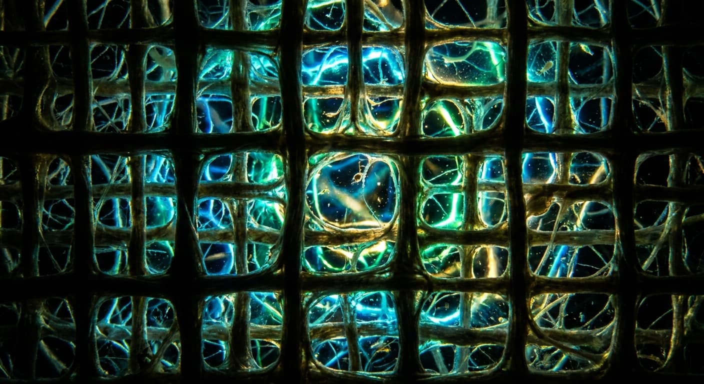
Have we missed a huge, unknown network in the body?
For centuries, human anatomy has been measured down to the smallest cell. But deep inside you - just under your skin and around your organs - there seems to be a gigantic, fluid-filled labyrinth that has remained completely invisible until now. A seemingly innocuous step in the way tissue is traditionally prepared for the microscope caused this complex network to collapse and become invisible before our eyes for decades. Only now is a far-reaching structure revealed that acts like a biological shock absorber - and perhaps holds the dark secret of how tumor cells can spread so quickly throughout the body. What exactly is hidden in these undiscovered cavities?

Your free account
Save interesting articles, track your reading progress and connect with other readers. Joining Pandragora is free.
Discover more with Premium
Start a subscription to get unlimited access to all articles.
Scientists have discovered a previously overlooked, fluid-filled network in your body that acts as a kind of “shock absorber” to protect almost all of your organs. This structure, which is part of the intercellular space, consists of a lattice-like network of stable collagen bundles through which fluid constantly flows. This space remained hidden for a long time because the fine cavities collapsed in on themselves during conventional tissue examinations and looked like solid connective tissue. You can find this system in many areas of your body, such as the skin, intestines and lungs. Because these spaces are directly connected to the lymphatic system, they have a significant impact on how swelling occurs or how quickly cancer cells travel through your body. With this new knowledge, many of the functions of your organs and the development of diseases can be completely reassessed.
An overlooked network in the human body
Imagine looking at a massive, impenetrable rock wall. You are absolutely certain that there is no way through there - until the tide comes. Suddenly, invisible cracks fill with water, and what just looked like solid stone turns out to be a complex system of branching underwater caves. This is exactly what medical research has done for centuries when looking inside your body. We thought we knew exactly how your organs and skin are built: made of dense, protective walls of connective tissue. But as clinical studies on living patients now show, we have overlooked a vast, fluid-filled dimension that runs through almost your entire body.
This discovery concerns the interstitium, the fluid-filled space between your cells and tissues. Until now, this space was thought of more like a moist environment for individual cells, but in reality it is one of the largest fluid reservoirs in your entire organism. It is a huge, connected network of macroscopically visible cavities that extends far beyond the previously known microscopic cell spaces. This structure is not a rare special case, but a fundamental construct that forms the submucosa - a layer of tissue directly under the mucous membrane - of your entire gastrointestinal tract, your urinary bladder and your bronchi. Even your dermis, the leather skin, and the fascia that surrounds your muscles consist of this previously unrecognized system.
Why didn't anyone notice this before? The mystery lies in the way we have studied tissue for generations. When anatomists take a tissue sample, it is usually chemically fixed and dehydrated so that it can be viewed under a microscope. At this moment the following happens: The liquid that covers these spaces like little balloons escapes. The structure collapses like an empty air shaft. All that remains are the dense collagen bundles, which now lie flat on top of each other and act like a solid wall of compact connective tissue. In a living body, what we have for decades referred to as “dense connective tissue” is actually an open, dynamic system of channels that is constantly being shaped by the rhythmic movements of the organs.
Only the use of the latest technology made this “aha moment” possible. With probe-based confocal laser endomicroscopy (pCLE), a technology that allows doctors to look into living tissue at a fixed depth of 60 to 70 micrometers during a mirroring, the invisible suddenly became visible. At a depth of 60 to 70 micrometers, what appeared was not a solid block, but a fascinating, web-like pattern of dark bands that enclose wide, bright spaces. This discovery forces us to reevaluate human anatomy. In many places, your body does not consist of dense barriers, but of a dynamic, fluid architecture that fundamentally changes our understanding of organ mechanics and explains how tumor cells can spread throughout the body via this network.
The error in tissue preservation
The decisive advance occurred during routine operations on the bile ducts between 2012 and 2013. To examine the delicate architecture of the tissue in real time, 13 patients received an injection of 2.5 milliliters of a 10 percent fluorescein solution into their bloodstream shortly before the surgical removal of tissue parts. This dye spreads quickly throughout the body and causes liquids to glow under laser light. What the researchers then observed using probe-based confocal laser endomicroscopy (pCLE) was astonishing: within 30 seconds of the infusion, the tissue lit up brightly at a depth of 60 to 70 micrometers. But it wasn't a uniform glow.
A clear, net-like pattern emerged in this fluid-filled space. It consisted of dark bands about 20 micrometers wide surrounding large, bright cavities like a grid. For comparison, the scientists also examined a control group that had not received any dye - the image remained dark there. But how could such a striking structure, which runs through almost the entire body, escape science for so long? The answer lies in an almost tragic irony of classical medicine: in order to protect and preserve tissue, we have unconsciously destroyed it.
Imagine a wet bath sponge brimming with water with its pores wide open. When you prepare this sponge for detailed examination in the laboratory, you traditionally press it flat, remove all moisture and fix it with chemicals so that it no longer changes. At the end you have a hard, solid block in front of you. Anyone who only sees this dry block would never imagine that the original object was a soft structure full of water-filled cavities. This is exactly what happens during standard tissue preparation in pathology. Drainage and fixation causes the fluid to escape from the interstitium, whereupon the supporting collagen bundles collapse and stick together tightly. The result under the microscope then looks like a solid wall of dense connective tissue.
In order to circumvent this effect and demonstrate the real structure in the laboratory, the researchers had to resort to a radical method: shock freezing. Immediately after removal, the tissue was frozen using cryopreservation before the fluid could escape. This was the only way the cavities remained open like small, frozen lakes. This ice fabric was then painstakingly cut in precise steps, each just 5 micrometers thick. Only these extremely thin layers revealed under the microscope exactly what had previously been seen in the living patients: a complex framework of fibers that created wide spaces for the tissue fluid. This finding proves that what we previously thought was a massive barrier in your submucosa is actually an open, fluid-filled canal system.
Asymmetrical walls and bare collagen
If you really want to understand the dimensions of this hidden space, take a look at medical imaging: ultrasound data shows that this network in your bile ducts makes up a remarkable 90 percent of the total wall thickness. So it's not a small gap, but the actual foundation of the tissue structure. Using a special technique that uses laser beams with a wavelength of 900 nanometers, the architecture of this room can be made visible like a glowing skeleton. This reveals a complex framework made up of thick collagen bundles - the fibers that give your body stability - as well as elastic fibers for the necessary flexibility.
However, the truly amazing thing only becomes apparent when the magnification reaches the nanoscale. In wafer-thin sections just 80 nanometers thick under the electron microscope, an anatomical peculiarity becomes visible that is found almost nowhere else in your body. Imagine a man-made river channel with a bank lined with protective tiles on only one side. On the opposite side, the bare earth lies completely unprotected and is washed directly around by the flowing water. This is exactly how the collagen bundles in this space are structured: they are asymmetrical. Only on one side are they covered by extremely flat, spindle-shaped cells, while the other side of the bundles is completely bare and is in direct contact with the circulating fluid.
The fact that this structure remained unrecognized for so long is also due to the fact that these cells could not really be assigned to any known category. They behave like molecular cross-border crossers. In laboratory studies it was found that although they carry certain markers such as CD34 and vimentin - proteins that are typically found on connective tissue cells or precursor cells - they lack one crucial property: they have no basement membrane. This is a thin separating layer that normally lies like a carpet under cover cells. Here, however, the cells adhere directly to the bare collagen without any intermediate layer.
One might assume that it is the lining of blood or lymph vessels, but the scientific evidence suggests otherwise. The cells lack all the typical features of a vessel's inner wall, such as the Weibel-Palade bodies - tiny storage organs for coagulation factors - or pinocytotic vesicles, which are responsible for substance transport. Tests for classic endothelial markers such as CD31, ERG or LYVE-1 were consistently negative. Smooth muscle actin, nuclear beta-catenin or the stem cell marker CD117 could not be detected either. Only the marker D2-40 was positive in the samples from the bile ducts, but was absent in all other tissues.
This discovery in the laboratory proves that you have a completely new system in front of you: a space in which liquid-filled cavities are directly bordered by naked protein matrices. This is highly physiologically relevant because collagen fibers are electrically charged molecules. The fact that they are directly exposed to the tissue fluid without a protective cell barrier could have far-reaching consequences for how cells in your immune system or even migrating pollutants interact with your connective tissue. It is an open, dynamic interface that is extremely rare in its kind in the human organism.
Shock absorbers and prelymphatic ducts
These cavities, lined on one side with cells, are not an anatomical curiosity of the bile ducts, but a fundamental structural principle of your body. You can find it wherever tissue is under mechanical stress and rhythmically contracts or stretches. It runs through the entire submucosal membrane of your gastrointestinal tract - from the esophagus to the stomach to the large intestine -, supports your urinary bladder, covers your bronchi and arteries and forms the deep layers of your dermis and the fascia of the musculoskeletal system.
The system forms a body-wide layer of interconnected, fluid-filled cavities that act as a network of shock absorbers. When you apply external pressure to your skin or when your intestines expand after a meal, this fluid-filled layer acts as a dynamic shock absorber. Since the collagen bundles form a stable grid that encloses the liquid, the tissue can flexibly absorb mechanical stress. The spaces are compressible and stretchable at the same time, which prevents your tissue from becoming rigid or brittle under constant mechanical stress.
But this “pop sheet” is more than just a protective cushion; it is a highly efficient wastewater system. It is a prelymphatic space - a kind of vestibule for the lymphatic system. Before the tissue fluid reaches the actual lymphatic vessels, it flows through this extensive labyrinth. The examination of four tattooed tissue samples from the colon shows that this system is functionally directly connected to your lymph nodes. The black pigments of the tattoo didn't just stay in place. Instead, phagocytes of the immune system, so-called macrophages, migrated into these interstitial cavities, absorbed the dye and traveled through the tunnel network to the draining lymph nodes.
Interestingly, in healthy tissue, these macrophages do not normally patrol these spaces. Clinical samples show that they only swarm this network massively when they have to react to foreign substances or stimuli. How important this reservoir is for fluid balance can be observed in the event of illness: in patients with incarcerated hernias, the fluid builds up in the submucosal membrane to such an extent that the protein-rich fluid - which appears pink in histological sections - massively pushes the collagen bundles apart. What was previously dismissed as non-specific swelling now turns out to be a measurable overload of this canal system.
In retrospect, traces of this network can even be found in the history of medicine. As early as 1906, the anatomist F.P. Mall the so-called “space of mall” in the portal region of the liver. His drawings, which were based on injection studies, show striking similarities to the structures we can see in the living body today thanks to state-of-the-art laser technology. It seems as if we have forgotten for a century what early researchers already suspected: your body is not a static structure of solid walls, but a highly dynamic reservoir in which fluid is constantly in motion to provide protection and remove waste.
The serious role in cancer and swelling
When this system works smoothly, it is a lifesaver for your organs. But these “subway tunnels” in your body have a dangerous downside: they can become an easily passable route for unwanted intruders to spread. What is intended as protective cushioning and an efficient disposal network can act like an open transport system for criminal gangs in the event of illness - in this case for cancer cells.
This risk becomes particularly clear when you look at cases of stomach cancer or stage T2 skin cancer where doctors have documented this phenomenon. A T2 tumor is a critical turning point: it has already invaded the submucosal membrane or the dermis, but has not yet reached the deeper muscle layers. In all five cases, the doctors found metastases in the draining lymph nodes, although the classic histological examination did not show any invasion of the cancer cells into blood or lymph vessels. The discovery of this fluid-filled space now provides the missing explanation. The tumor cells simply have to gain access to this extensive network of tunnels. Once there, they don't even have to move around under their own power. The mechanical pressure of your body, such as the rhythmic peristalsis of your intestines or the compression of your skin when you move, drives the cells like an underground conveyor belt straight to the next “station” - the lymph node.
This knowledge also sheds completely new light on fluid displacement into third space, a phenomenon that often occurs in medicine after major operations or organ failure. For example, if lymph nodes have been removed after cancer surgery or if the liver, kidney or heart fails, massive swelling, so-called edema, can occur. This fluid doesn't simply disappear into the tissue; it floods and expands precisely this interstitial canal system. The typical edema, which develops within a very short time when the bile ducts are blocked, also takes place directly in this network. It's as if a sewer pipe were to clog and flood the entire connected tunnel system.
Beyond pure transport, the cells that delimit these spaces could act as a kind of “first responder” in the event of inflammation and scarring. There are initial indications that the flat, spindle-shaped cells in these cavities are the precursors of the cells that, in the event of chronic irritation, trigger massive tissue hardening, a so-called sclerosis. This could explain why diseases such as primary sclerosing cholangitis, biliary atresia or chronic inflammatory bowel disease leave such profound and often irreversible marks on your connective tissue.
This system even seems to play an infamous role in keloids, those bulging scars that often grow painfully beyond the original wound area. In such scar tissue, collagen-rich bundles and greatly enlarged cavities have been observed, appearing like an exaggerated, misguided version of the normal interstitial spaces. Since keloids tend to occur in places with high mechanical stress, researchers suspect a direct connection between the physical forces, the flow of fluid in these tunnels and the incorrect response of the cells resident there. Your body not only uses this network for maintenance, but also reacts to massive stress via this system - sometimes with serious consequences for the tissue structure.
The open future of anatomical research
With these discoveries, medical research has literally turned on the lights on a completely unknown floor of our own home. For a long time we thought that the foundation of your body was made of solid stone, but now we are looking into an extensive system of corridors and water pipes. We now know they are there, and we are beginning to understand how the water flows through the pipes - but we still have no idea who the actual residents of this floor are or what rules they live by there.
The biggest gap in our knowledge concerns the enigmatic cross-border cells that line the collagen skeleton. It is completely unclear whether these are a completely new type of connective tissue cells, so-called fibroblasts, or even mesenchymal stem cells - biological all-rounders that can transform into different types of tissue. We do not yet know whether these cells themselves are the ones that produce and maintain the massive collagen bundles. If they are indeed the architects of this space, they could hold the key to how your body heals wounds or why it scars with age and illness.
Another physical mystery is the “naked” side of the collagen bundles. Since they do not have a protective cell layer there, they come into direct contact with the flowing tissue fluid. Because collagen fibers are electrically charged molecules, they form a highly efficient, physiologically active surface. Whether and how the cells of your immune system that patrol these tunnels interact with this bare collagen is one of the most burning questions in current research. It could be that biochemical signals are being transmitted here that we have never even dreamed of before.
This discovery also opens up a whole new perspective on how your body exchanges information. Since the fluid in the submucosal membrane of your intestine is probably driven by rhythmic peristalsis, an exciting hypothesis arises: Immunological or hormonal signals could migrate in this network in a strictly directed manner - always from top to bottom, just like the chyme inside the intestine. This would mean that one section of your body could tell the next part “downstream” via the interstitium what to expect.
In the future, this knowledge could revolutionize the way we detect disease. If this network is indeed a body-wide highway for fluids and cells, then the interstitium is a gold mine of information. Researchers are considering that directly aspirating and analyzing this interstitial fluid could become a powerful diagnostic tool. Instead of waiting for markers to show up in the blood, doctors could look for the first signs of cancer or inflammation in this wet tunnel system. We have only just begun to read the blueprints of this hidden system, and each new room we enter could show us how to better protect and heal our bodies.
──────────────────────────────────────────────────
Those
Benias, P. C. et al., 2018, Structure and Distribution of an Unrecognized Interstitium in Human Tissues, DOI: 10.1038/s41598-018-23062-6
──────────────────────────────────────────────────
Share or save this article
Share this post or save it for later. A like helps make relevant content more visible.
We place great emphasis on accuracy in our research. Nevertheless, errors can occur. We also recommend reading the corresponding primary literature listed in the sources. The content on Pandragora is for general information only and does not replace professional medical advice or treatment. Use of the information provided is expressly at your own risk and responsibility; any liability is excluded.